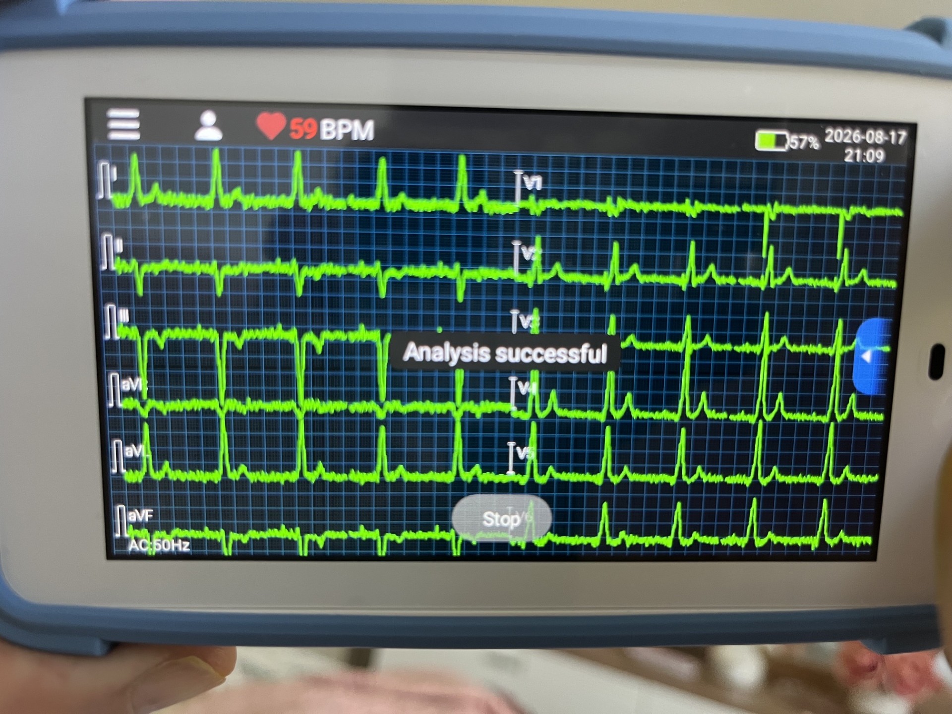
The reading put forward
V6 is encouraging; V1 is inconclusive and probably technically unreliable.
V1: The complexes change markedly during the recording — from very small signals to deep predominantly negative rS/QS complexes. That variation suggests electrode contact, placement, automatic gain or stabilisation artefact. I do not see a consistent terminal positive R/R′, which is commonly expected with successful LBB-area activation. This makes V1 less typical, but it does not demonstrate loss of LBB capture.
V6: There is a stable, dominant R wave followed by a small but definite terminal S wave. That morphology is compatible with LBBAP and resembles the lateral terminal-S feature previously observed. The rapid-looking rise of the R wave is also encouraging.
The short answer
The direction of that reading is defensible, but it is over-confident about V6 and under-confident about a more basic problem: nothing in this tracing establishes that these are paced beats at all. Once that is unresolved, "encouraging" and "inconclusive" are not two different verdicts — they are the same verdict applied to two leads.
1. The premise that is not anchored
No pacing artefact is identifiable anywhere in the image with any confidence. A rate of 59 bpm sits exactly where a programmed base rate could plausibly live, which is suggestive but not evidential. Many handheld and consumer-grade recorders apply anti-aliasing and mains filtering, and sample at rates, that attenuate or entirely drop the narrow stimulus artefact. So the absence of visible spikes is not evidence of absent pacing — but neither is it evidence of pacing.
This matters because every established surface criterion for left bundle branch area capture is measured from the stimulus:
- V6 R-wave peak time (V6RWPT / Stim-LVAT) — from stimulus onset to the peak of the R wave in V6. No stimulus, no start point.
- The V6–V1 interpeak interval — the separation between R-wave peaks in the two leads, both referenced to the same stimulus. Same problem.
- Output-dependent transition — the abrupt shortening or lengthening of those intervals as output is decremented, which is the most robust of the three. It requires a deliberate programming manoeuvre that was never performed here.
There is a fourth consequence that is easy to overlook: without a stimulus marker, paced and intrinsic complexes cannot be separated. A tracing may be describing conducted beats while being read as though it described paced ones.
The core point
Morphology criteria in conduction system pacing are timing criteria wearing the clothes of shape criteria. Strip out the stimulus and what remains is a description, not a measurement.
2. Why the V6 observation does not discriminate
A stable dominant R wave with a small terminal s in V6 is a real and correctly described finding. The difficulty is what it rules out, which is close to nothing.
That morphology is also the ordinary intrinsic lateral morphology of a normally conducted beat. It is what you would expect to see in a person with intact left-sided conduction and no pacing whatsoever. It is therefore equally compatible with:
- intrinsic conduction with no ventricular pacing;
- non-selective left bundle branch capture;
- selective left bundle branch capture;
- left ventricular septal myocardial capture without engagement of the conduction system.
"Compatible with LBBAP" is true and nearly vacuous. The clinically live question is not could this be LBBAP but is this conduction system capture or septal myocardial capture — and V6 shape alone does not touch it. The discriminator is timing: a short V6 R-wave peak time measured from the stimulus, and more reliably its abrupt change at output decrement.
"The rapid-looking rise of the R wave is also encouraging" is where the reasoning quietly substitutes an impression for a measurement. R-wave peak time is a number in milliseconds. On a screen photograph, at this sweep speed, with the interface partially occluding the V6 onset, it is not obtainable — and a visual impression of steepness is strongly influenced by amplitude scaling, which is exactly what the same reading flags as unreliable one paragraph earlier in V1.
3. V1 — right conclusion, different mechanism
The variation is probably acquisition, not gain
The jump from tiny complexes to deep rS/QS morphology across the trace is better explained by sequential precordial acquisition than by automatic gain or baseline stabilisation. Many handheld six-electrode systems do not capture V1–V6 simultaneously; a single chest electrode is moved from position to position during the recording, and the display fills in each precordial channel as it is acquired.
If that is what happened, some of the complexes shown under the V1 label may not be V1 at all. That is a different and more serious problem than gain artefact, because gain artefact distorts a real lead while repositioning replaces it.
The caveat about terminal R/R′ is correct, and worth stating more strongly
The absence of a consistent terminal positive deflection in V1 does not exclude left bundle branch capture. It can be absent when:
- the V1 electrode is placed higher than the fourth intercostal space — a very common error, and unverifiable on a photograph;
- capture is left ventricular septal rather than conduction system, in which case delayed right ventricular activation may be less pronounced;
- individual anatomy, chest geometry or cardiac rotation attenuates the terminal vector.
The converse is equally true and less often said: a qR or rSR′ pattern in V1 is not specific for conduction system capture. Right ventricular septal capture can produce it. V1 is a supporting lead in this assessment, not an adjudicating one, in either direction.
4. Signal quality issues to correct before repeating
- The AC filter is set to 50 Hz. If the recording was made on a 60 Hz mains supply, the notch is sitting on the wrong frequency and mains interference passes through unfiltered. That plausibly accounts for a substantial share of the baseline noise.
- The rectangular deflections in III and aVL are not physiology. Excursions with vertical edges and flat tops, confined to particular channels, are amplifier saturation or lead-off events. They are a hardware state, not a cardiac signal.
- Interface elements occlude the data. The "Analysis successful" toast covers part of V3, and the Stop control sits over the V6 onset — precisely the region needed for any R-wave peak time assessment.
- "Analysis successful" is not a quality certificate. It reports that the algorithm ran to completion, not that the signal was adequate or the interpretation correct.
- A photograph of a screen is not the recording. It adds moiré, perspective distortion and compression loss on top of everything above. If the device exports a PDF or a raw file, that is what should be reviewed.
5. What each claim is actually worth
| Claim in the proposed reading | Verdict | Reason |
|---|---|---|
| V6 morphology is compatible with LBBAP | True but non-discriminating | It is also normal intrinsic lateral morphology, and also compatible with LV septal capture |
| The R-wave rise in V6 is rapid | Not established | An impression substituting for a millisecond measurement that cannot be made from this image |
| V1 is technically unreliable | Agreed | Though the likelier mechanism is sequential precordial acquisition, not gain artefact |
| Absent terminal R/R′ in V1 does not prove loss of capture | Correct | Electrode height, septal capture and anatomy all suppress it; the converse is also non-specific |
| Overall: capture is probably intact | Not supported either way | No stimulus is identifiable, so paced and intrinsic beats cannot be separated |
6. What would actually answer the question
- A diagnostic-quality twelve-lead with visible stimuli, recorded on a machine that preserves the pacing artefact, with verified electrode placement.
- Programmed output decrement during that recording, watching for an abrupt transition in QRS morphology and in stimulus-to-V6 peak time at the point where conduction system capture is lost but myocardial capture persists.
- Device interrogation: the separate capture thresholds for the two capture types, unipolar and bipolar impedance, sensed and paced electrogram morphology, percentage of ventricular pacing, and atrial arrhythmia burden.
That is a device clinic procedure. No handheld tracing, however clean, substitutes for it — and a clean tracing cannot retroactively supply the output decrement step that was never performed.
Bottom line
This tracing supports "no alarming finding is visible" and nothing stronger. It is not evidence for preserved left bundle branch capture, and it is not evidence against it. The honest position is that the recording does not carry the information the question requires — which is a more useful conclusion than a confident one, because it points at the right next step rather than closing the file.
Related
- Can a noisy handheld twelve-lead ECG be interpreted in an LBBAP patient?
- What device interrogation adds beyond symptoms and the surface ECG
- Persistent left/superior QRS axis after LBBAP
References
- Jastrzębski M, et al. Left bundle branch area pacing outcomes: the multicentre European MELOS study. Europace. 2022. PMID: 35979977
- Jastrzębski M, et al. The V6–V1 interpeak interval: a novel criterion for the diagnosis of left bundle branch capture. Europace. 2022. PMID: 34718539
- Wu S, et al. Criteria for confirming left bundle branch capture during left bundle branch area pacing. Heart Rhythm. 2021. PMID: 33677102
- Burri H, et al. EHRA clinical consensus statement on conduction system pacing implantation. Europace. 2023. PMID: 37262189
- Kligfield P, et al. Recommendations for the standardization and interpretation of the electrocardiogram. Circulation. 2007. PMID: 17322457