LBBAP Pacemaker Incision Healing: 2 Weeks vs 7 Weeks Post-Procedure
Reviewed by the Artificial Intelligence Medical Team
Advertisement
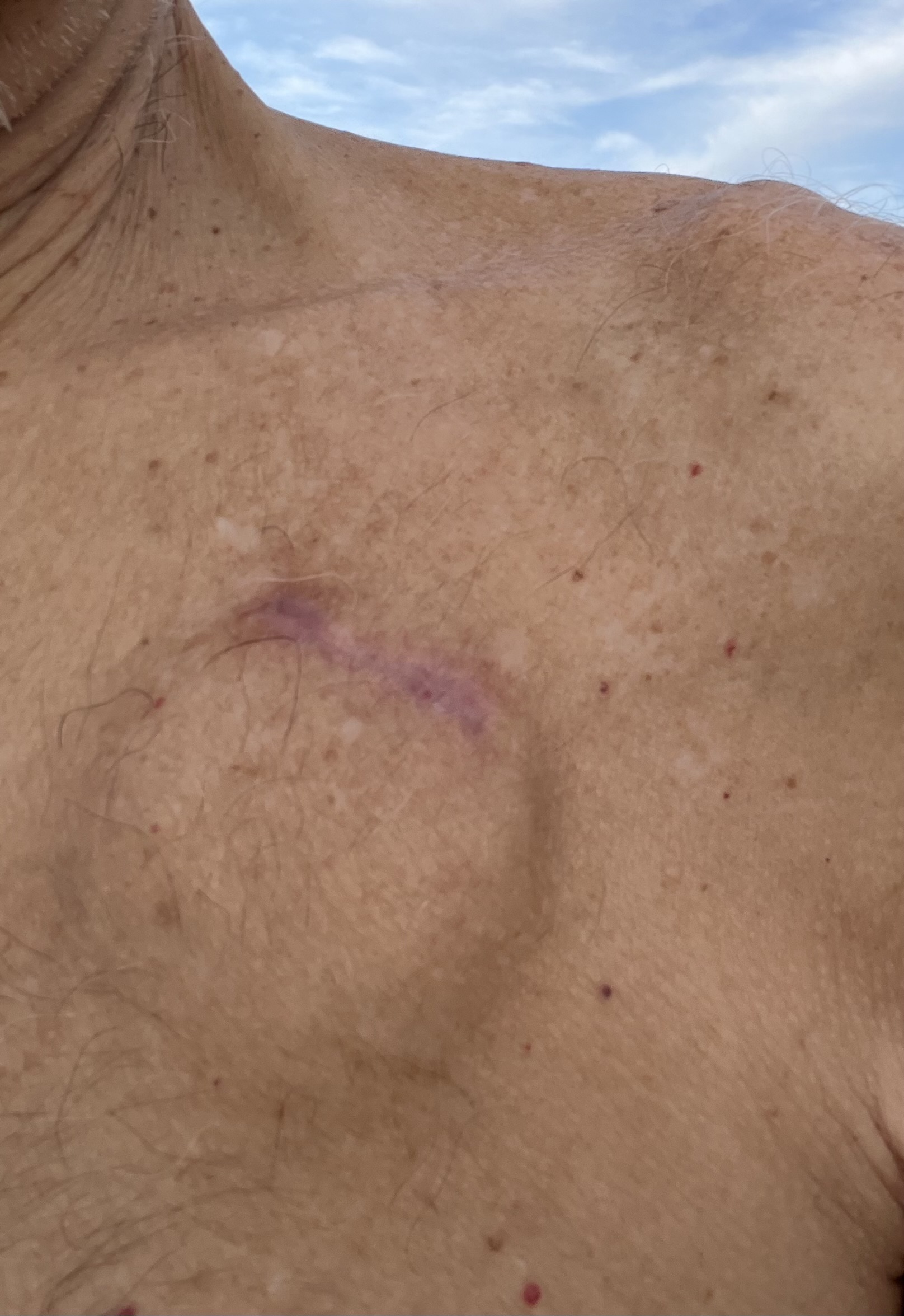
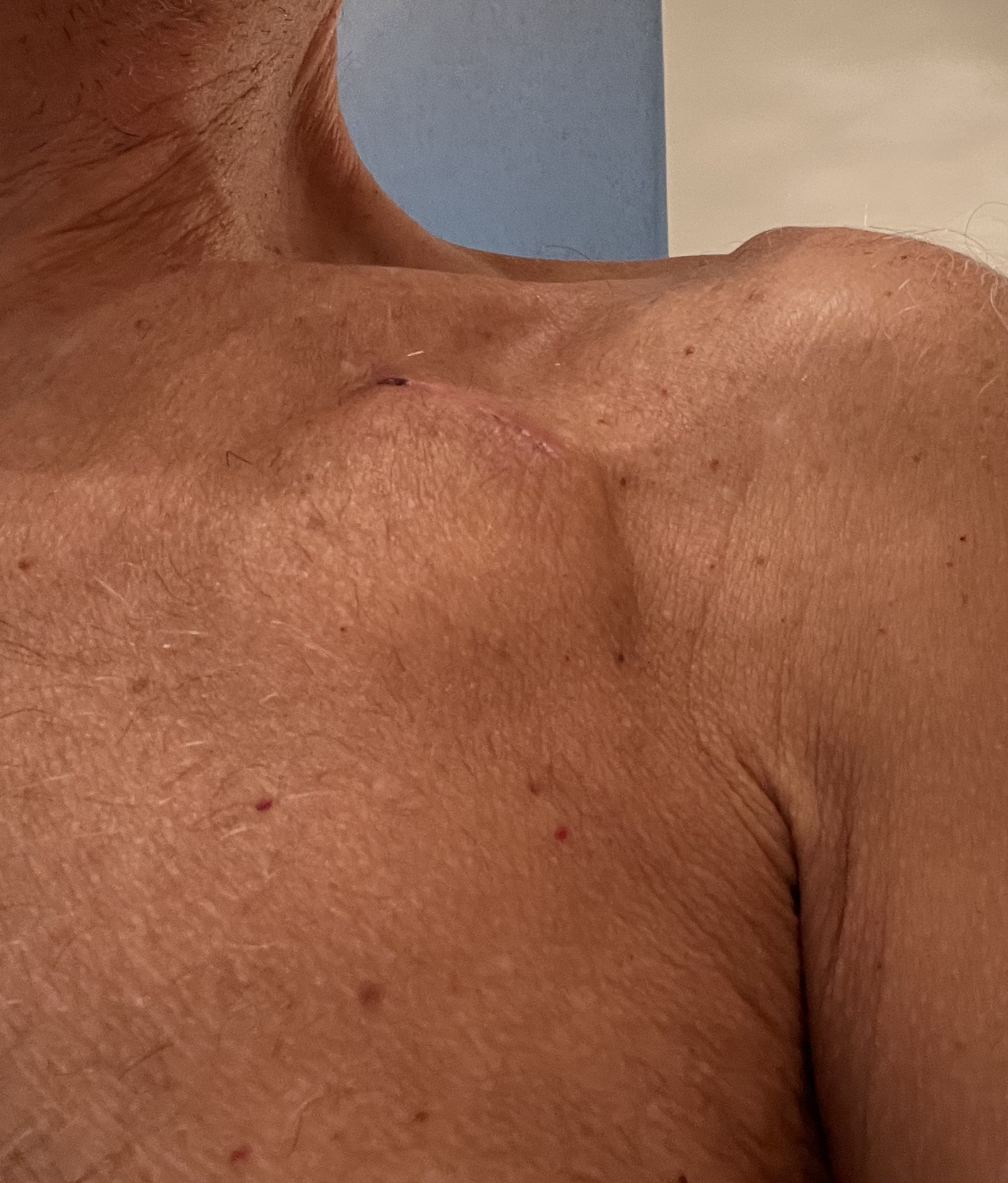
Image 1 — LBBAP pocket incision at 7 weeksImage 2 — LBBAP pocket incision at 2 weeks
Question: Can you please give your opinion on these two pictures showing an LBBAP pacemaker 2 weeks after the procedure and 7 weeks after the procedure?
Comparing the two timepoints
2 weeks (Image 2): Incision line still visible with a residual suture mark/scab at the medial end, surrounding erythema, and a visible linear healing ridge along the infraclavicular incision. Pocket area shows mild edema/contour change consistent with early post-operative healing.
7 weeks (Image 1): The linear scar has matured considerably — color has shifted from pink/erythematous to a violaceous/purple hue typical of a scar in the early remodeling phase (collagen deposition still active, vascularity not yet fully regressed). The scar is less raised, the suture mark/scab from week 2 has resolved, and the surrounding skin appears free of induration, fluctuance, or erythema spreading beyond the incision line — no signs of pocket infection or hematoma.
Advertisement
Overall assessment
The overall trajectory looks like normal expected healing: progression from a fresh, slightly inflamed incision to a maturing linear scar with the violaceous discoloration that is typical at this stage. This usually continues to fade toward a flatter, paler scar over the following months.
The scattered red puncta visible in both images appear to be cherry angiomas or petechiae unrelated to the pocket itself — common incidental skin findings, not a device-related concern.
There are no signs of dehiscence, erosion, pocket hematoma, or device migration in either image based on the external appearance alone.
Advertisement
This assessment is based solely on external photographic appearance and is provided for general educational purposes. It is not a substitute for in-person evaluation by the implanting team, which remains essential for any concern regarding infection, dehiscence, erosion, or hematoma.